Upper Leg Tendon Anatomy - Human Anatomy Leg Tendons | Leg muscles diagram, Leg ... / Tendons of the anterior compartment of the leg, the anterior tibial vessels, and the deep peroneal nerve pass under it.
Upper Leg Tendon Anatomy - Human Anatomy Leg Tendons | Leg muscles diagram, Leg ... / Tendons of the anterior compartment of the leg, the anterior tibial vessels, and the deep peroneal nerve pass under it.. And it is also critical to the walking process. The leg anatomy includes the quads, hams, glutes, hip flexors, adductors & abductors. The appendicular skeleton includes the bones of the shoulder girdle, the upper limbs, the pelvic girdle, and the lower limbs. They are innervated by the tibial nerve, a terminal branch of the sciatic nerve. Collectively, the muscles in this area plantarflex and invert the foot.
Information on the central tendon of the diaphragm by the anatomyzone daily feed. Look for subcutaneous landmarks to figure out where the bones go. The large achilles tendon is the most important tendon for walking, running, and jumping. Tendons are fibrous cords attached to muscles and bone. Try to do both of these exercise on your own, before i post my answer briefly, it sits inside the quadriceps tendon and connects it to the front of the tibia by way of the patellar ligament.
Fitness for You: Fitness for You - Lower Body Exercises from 3.bp.blogspot.com Leg anatomy anatomy poses anatomy study anatomy art anatomy drawing human anatomy anatomy images body reference anatomy anatomical drawings sketchbook ,artist study resources for art students with thanks to artist simone bianchi, how to draw the human figure. Try to do both of these exercise on your own, before i post my answer briefly, it sits inside the quadriceps tendon and connects it to the front of the tibia by way of the patellar ligament. Learn the origin/insertion, functions & exercises for the leg rotating your upper leg and pelvis to the inside or outside of your body's center line. The leg is composed of five distinct sections: The lower leg is comprised of two bones, the tibia and the smaller fibula. The tendons that control movement in your hands, wrists and fingers run through your forearm. In human anatomy, the lower leg is that part of the lower limb that lies between the ankle and the knee. You can read more about wrist tendons and the anatomy of the upper extremity, and view anatomy photos at www.handcare.org.
Tendinous sheath of right flexor pollicis longus radial bursa.
These originate from the outer lower leg bone (fibula) then travel down along the outside of your leg where they insert into various bones in. Try to do both of these exercise on your own, before i post my answer briefly, it sits inside the quadriceps tendon and connects it to the front of the tibia by way of the patellar ligament. Look for subcutaneous landmarks to figure out where the bones go. 935 x 1601 jpeg 153 кб. The appendicular skeleton includes the bones of the shoulder girdle, the upper limbs, the pelvic girdle, and the lower limbs. Upper legs anatomy — stock image. Do anatomy tracings over those to find the leg bones. The tendons that control movement in your hands, wrists and fingers run through your forearm. Hands are outstretched, holding onto the handles of the bench. Collectively, the muscles in this area plantarflex and invert the foot. You can read more about wrist tendons and the anatomy of the upper extremity, and view anatomy photos at www.handcare.org. Synovial tendon sheaths of right fingers. The upper leg begins at the hip and continues down to the knee.
It is the largest tendon of the parts of leg. Extends leg at knee in quad group. Try this movement out by standing on one foot with the other leg. The lower leg is comprised of two bones, the tibia and the smaller fibula. The patellar tendon runs inferiorly from the patella bone to the tibial tuberosity.
Anterior view of leg muscles from www.anatomynote.com Spicermanyt at checkout for 40% off this tutorial! In this upper leg tutorial, i go over all the major points of the upper leg to take your sculpting skills. Tendinous sheath of right flexor pollicis longus radial bursa. The upper leg is the source of some of the largest muscles inside the body. Learn the origin/insertion, functions & exercises for the leg rotating your upper leg and pelvis to the inside or outside of your body's center line. Use the mouse scroll wheel to move the images up and down alternatively use the tiny arrows (>>) on both side of the image to move the images. It blends with the fibrous pericardium above, helping to. There are several muscles which lie on the outside of your lower leg and are collectively known as the peroneal muscles (figure 1).
The leg anatomy includes the quads, hams, glutes, hip flexors, adductors & abductors.
Learn the origin/insertion, functions & exercises for the leg rotating your upper leg and pelvis to the inside or outside of your body's center line. They are innervated by the tibial nerve, a terminal branch of the sciatic nerve. Learn vocabulary, terms and more with flashcards, games and other study tools. Upper leg, knee, lower leg, ankle, and foot. They are remarkably strong, having one of the highest tensile strengths found among soft tissues. Use the mouse scroll wheel to move the images up and down alternatively use the tiny arrows (>>) on both side of the image to move the images. The nerve signals in these reflexes come from stretch receptors located in the joints, ligaments reflexes help to maintain proper muscle tone, balance, and responsiveness of the legs and feet to stimuli such as stepping on a sharp object. Leg anatomy anatomy poses anatomy study anatomy art anatomy drawing human anatomy anatomy images body reference anatomy anatomical drawings sketchbook ,artist study resources for art students with thanks to artist simone bianchi, how to draw the human figure. However, many reflex pathways are also active in the legs and foot. The human leg, in the general word sense, is the entire lower limb of the human body, including the foot, thigh and even the hip or gluteal region. Tendinous sheath of right flexor pollicis longus radial bursa. Synovial tendon sheaths of right fingers. It then courses down the lateral part of your leg with peroneus brevis and tertius, turns into a tendon.
The tendons that control movement in your hands, wrists and fingers run through your forearm. 1074 x 1856 jpeg 989 кб. To describe the mechanical properties of tendons. Anatomical structures and specific regions are visible as dynamic labeled images. There is no real division between the core and the upper leg;
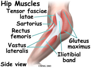
Ligaments, tendons, and muscles of the hip joint | Naples ... from www.zehrcenter.com The anatomical basis of clinical practice. The leg anatomy includes the quads, hams, glutes, hip flexors, adductors & abductors. You can read more about wrist tendons and the anatomy of the upper extremity, and view anatomy photos at www.handcare.org. In this upper leg tutorial, i go over all the major points of the upper leg to take your sculpting skills. Iliotibial band syndrome description the iliotibial band is the tendon attachment of hip muscles into the upper leg (tibia) just below the knee to the outer side of the front of the leg. Try this movement out by standing on one foot with the other leg. The thigh bone, or femur, is the large upper leg bone that connects the lower leg bones (knee joint) to the pelvic bone (hip joint). Synovial tendon sheaths of right fingers.
The thigh and leg bones articulate at the knee joint that is protected and enhanced by the patella bone that supports the quadriceps tendon.
Iliotibial band syndrome description the iliotibial band is the tendon attachment of hip muscles into the upper leg (tibia) just below the knee to the outer side of the front of the leg. Subscribe to learn interesting facts about the human body every day. The nerve signals in these reflexes come from stretch receptors located in the joints, ligaments reflexes help to maintain proper muscle tone, balance, and responsiveness of the legs and feet to stimuli such as stepping on a sharp object. Hands are outstretched, holding onto the handles of the bench. 1074 x 1856 jpeg 989 кб. Tendons of the anterior compartment of the leg, the anterior tibial vessels, and the deep peroneal nerve pass under it. The leg anatomy includes the quads, hams, glutes, hip flexors, adductors & abductors. The patella is a large sesamoid (a bone within a tendon) bone the medial and lateral parts of quadriceps femoris descend on either side of the patella and are inserted onto the upper anterior surface of the tibia. By spicer mcleroy in tutorials. In human anatomy, the lower leg is that part of the lower limb that lies between the ankle and the knee. This mri wrist coronal cross sectional anatomy tool is absolutely free to use. The leg is composed of five distinct sections: Learn the origin/insertion, functions & exercises for the leg rotating your upper leg and pelvis to the inside or outside of your body's center line.


0 Comments